
Mechanism of Injury
Lateral Tibial Plateau Fracture
An illustrated figure demonstrating the mechanism of injury of Jane Doe, completed in a medical legal style.
Roles: Research, Maquette creation, Sketch, Final rendering, Compositing, Editing
Audience: Judges, Jury, Lawyers
Clients: Professor Stephen Mader
Communication Objective: Illustrate the mechanism of injury for Jane Doe based on real radiological and case information
Medium: Web, Digital
Software: Procreate, Adobe Illustrator
Process
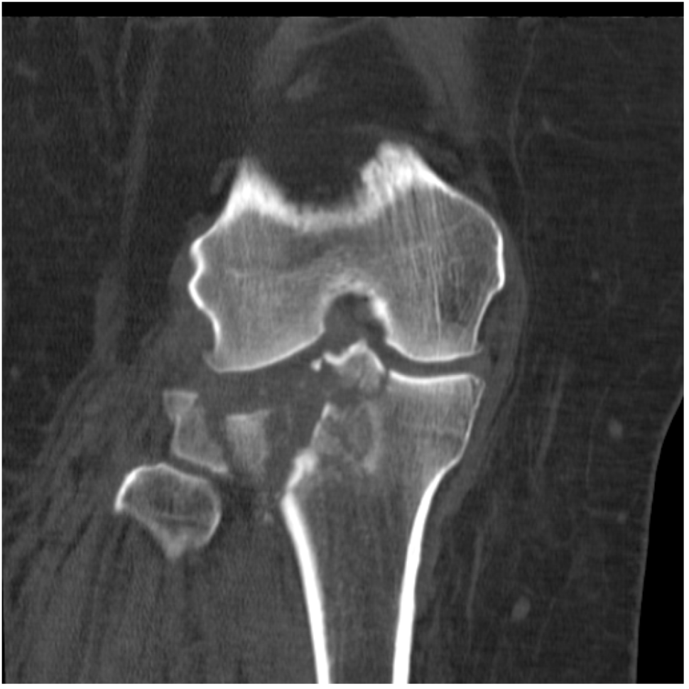
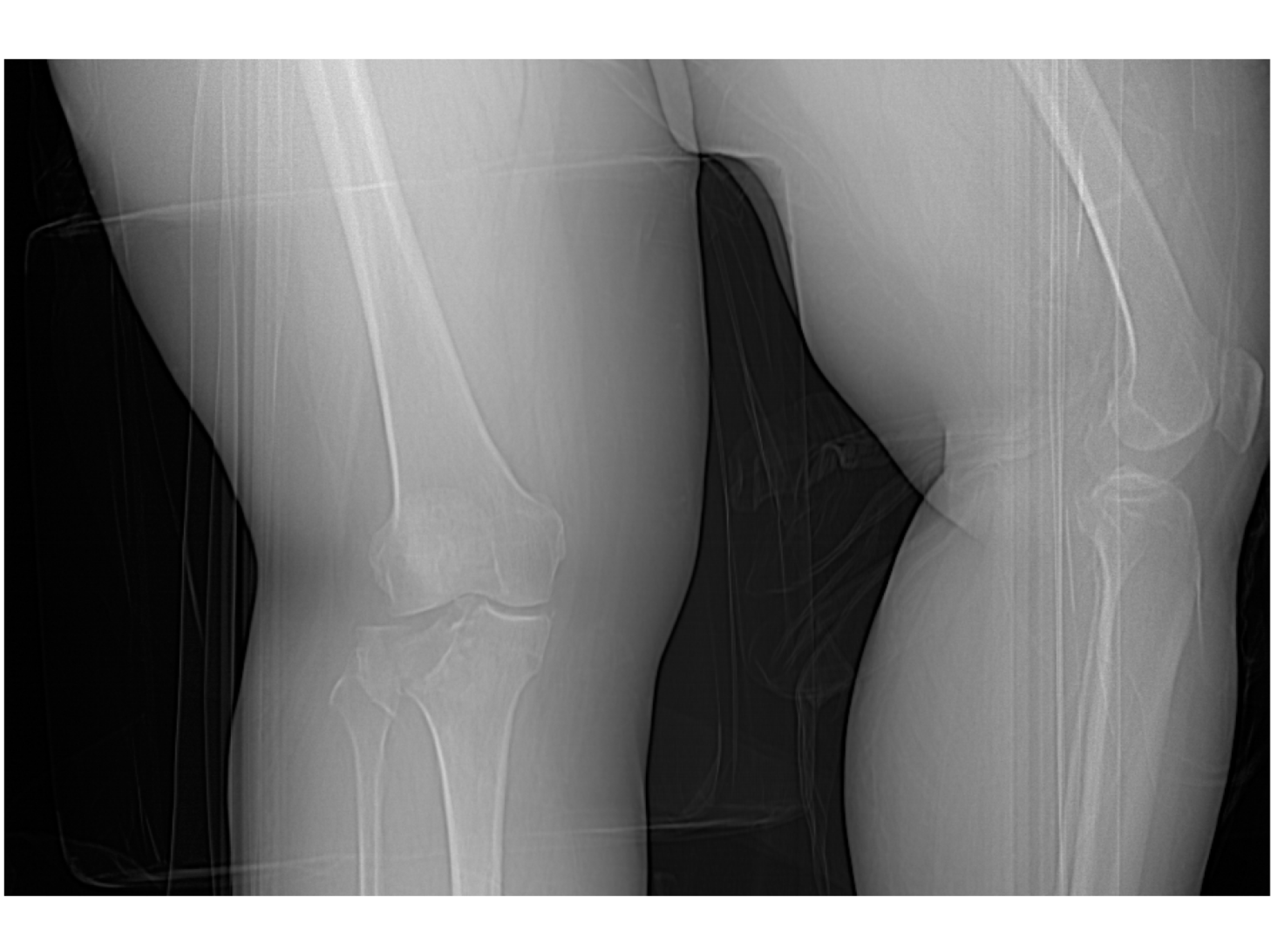
I was provided case files and radiological imaging of a real medical legal case (with permission) and began analyzing and parsing through the information to glean relevant details for the illustration such as how an injury occured, who was involved, what obstacles/environmental elements were involved, the forces and injury mechanics, and the details of the injured person.
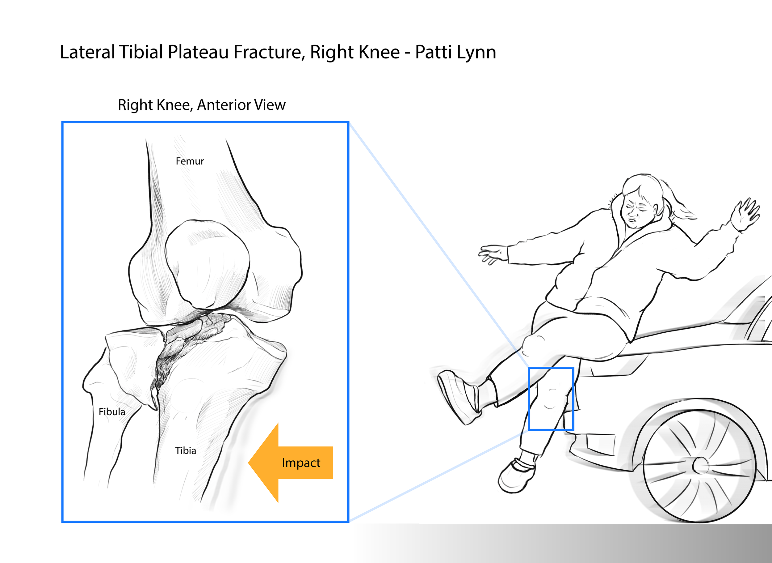
I enlisted the help of a classmate to get a reference image for the pose of Jane Doe as they collided with the vehicle.
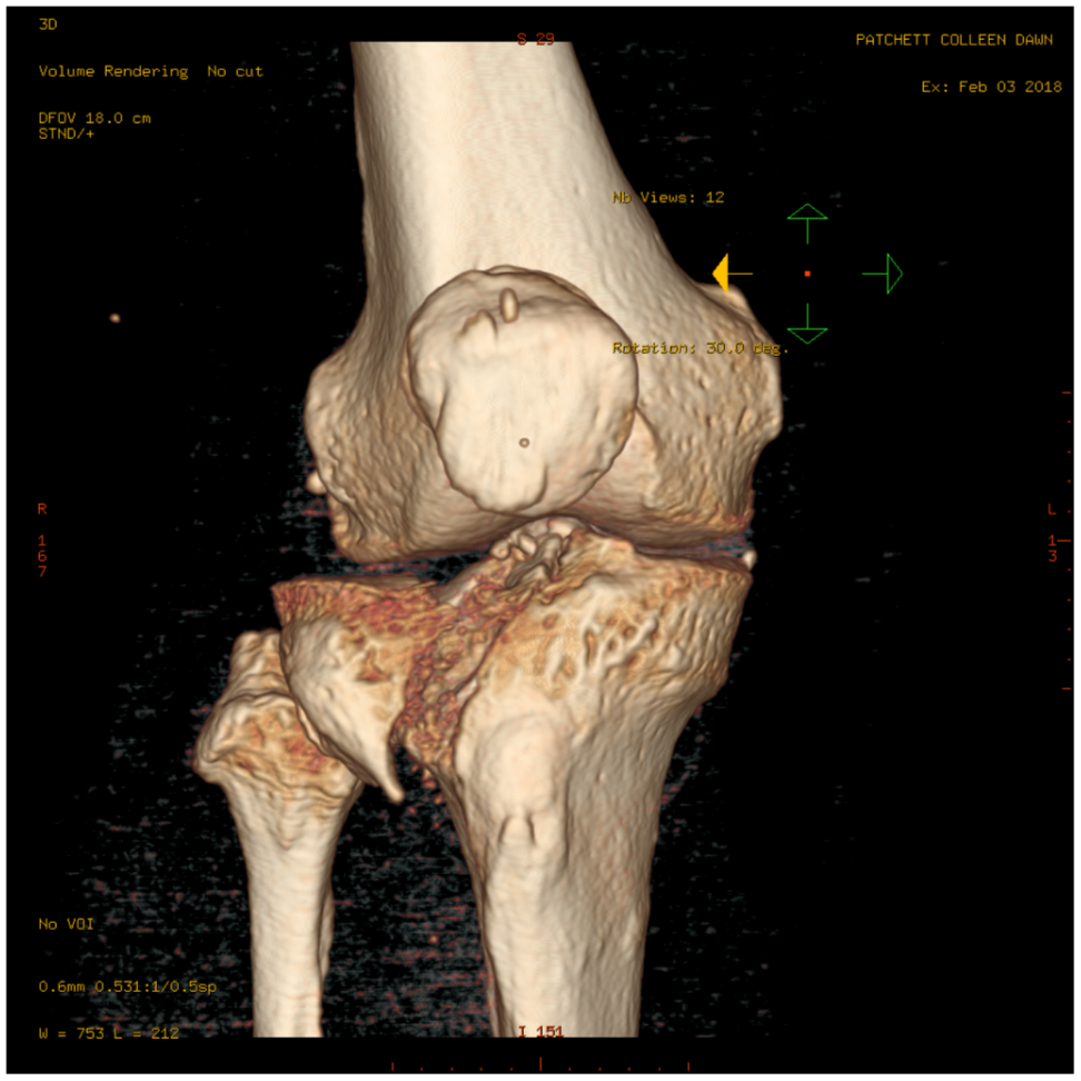
I determined the fracture to be a type II tibial plateau fracture, which was a result of the lower energy impact of a vehicle on Jane Doe’s medial aspect of their right knee.

A rough sketch was made.

After doing more research and receiving feedback, I refined the illustration.
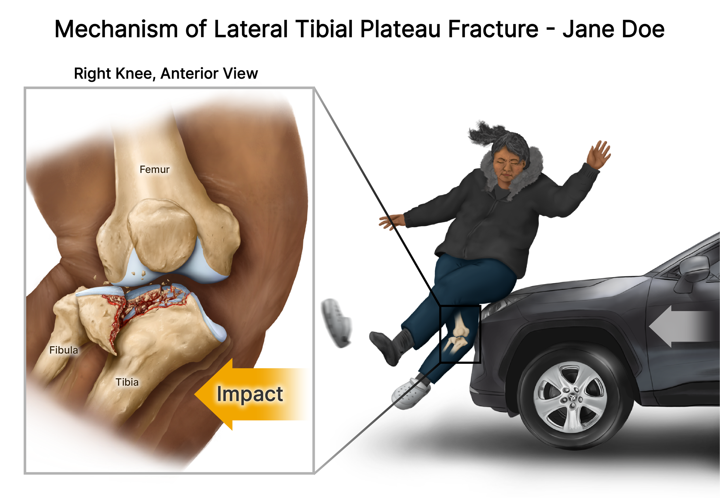
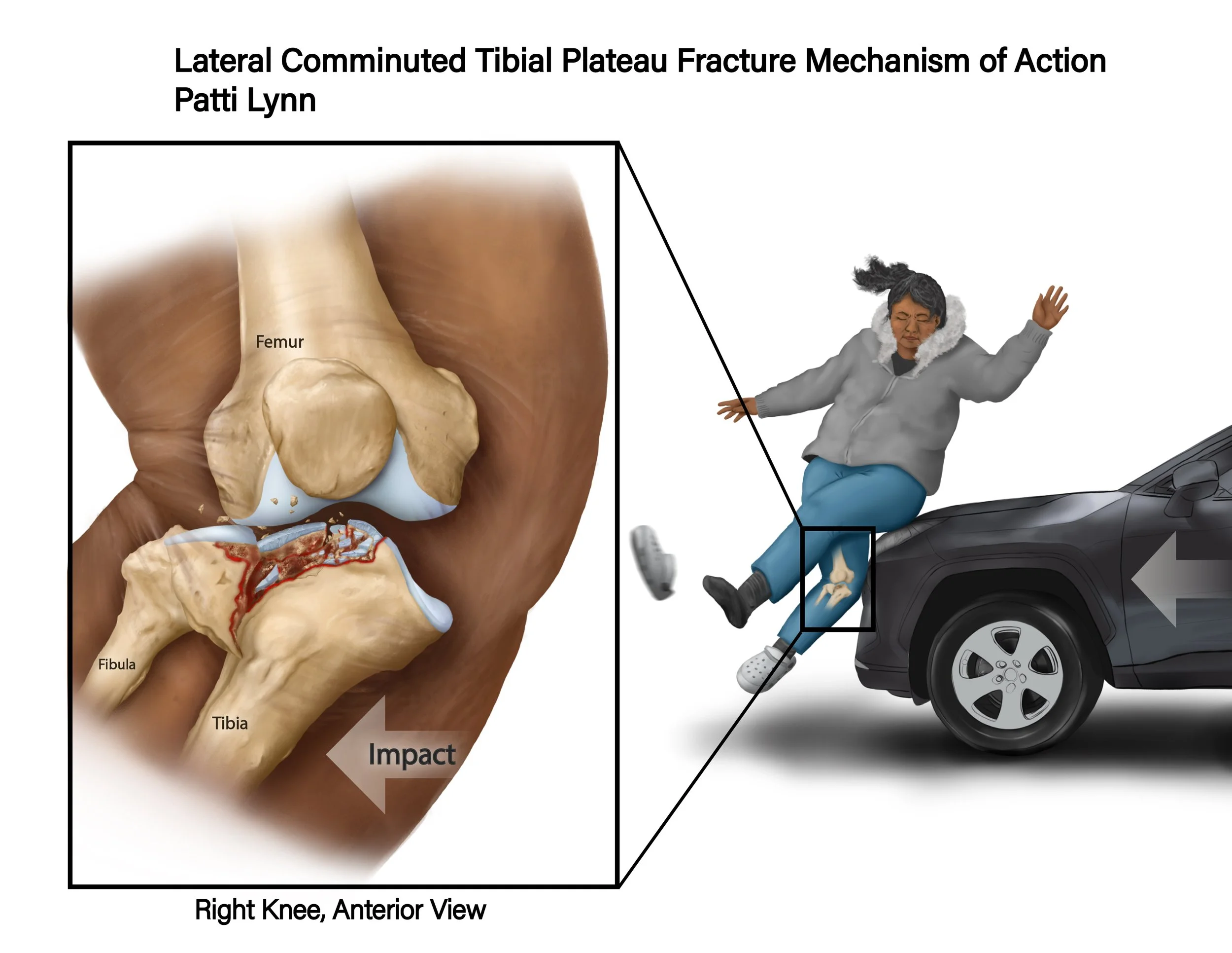
The final Illustration:
References
Agur, A. M. R., & Dalley, A. F. (2021). Grant's Atlas of Anatomy. Wolters Kluwer.
Platzer, W., & Shiozawa-Bayer, T. (2021). Color Atlas of Human Anatomy Vol 1. Locomotor System (5th ed., Vol. 1). Thieme.